
Gram Reaction Of Mycobacterium Tuberculosis
So, there I was, elbow-deep in… well, let's just say it wasn't a baking soda volcano experiment. Picture this: a dimly lit lab, the faint smell of something vaguely antiseptic, and me, squinting through a microscope at what looked like a chaotic smear of tiny, reddish-brown specks. My professor, a man whose tweed jacket seemed to have absorbed decades of scientific curiosity (and possibly some spilled coffee), peered over my shoulder. "Ah," he declared with a sage nod, "the notorious Mycobacterium tuberculosis. Quite the character, wouldn't you say?"
I, at that moment, mostly thought it was a character that was stubbornly refusing to behave. You see, my task was to perform a Gram stain. Simple, right? Dip, stain, rinse, repeat. Except, for Mycobacterium tuberculosis, it's more like a defiant shrug and a "nah, not playing by your rules." It’s a bit like trying to get a stubborn toddler to eat their veggies – they just have their own agenda.
And that, my friends, is how I learned that not all bacteria are created equal, especially when it comes to the seemingly straightforward Gram stain. This little anecdote, while slightly dramatized for your entertainment (okay, maybe only slightly!), perfectly sets the stage for our deep dive into the gram reaction of Mycobacterium tuberculosis. Because, let me tell you, it’s not what you might expect.
The Gram Stain: A Quick Recap (Because We All Need One, Right?)
Before we get into the rebellious nature of our favorite lung-invading microbe, let's quickly revisit what a Gram stain is all about. It’s basically the CSI of the microbiology world. You take a sample, you apply a series of stains and reagents, and you see what color the bacteria turn. Easy peasy, lemon squeezy… usually.
The magic (or the madness, depending on your perspective) lies in the bacterial cell wall. Gram-positive bacteria have a thick layer of peptidoglycan, which acts like a sponge. When you apply the primary stain (crystal violet) and then a decolorizing agent (like alcohol), this thick peptidoglycan layer traps the crystal violet, making the bacteria appear purple.
Gram-negative bacteria, on the other hand, have a much thinner peptidoglycan layer and an outer membrane. When the decolorizing agent hits them, it strips away the crystal violet. Then, you apply a counterstain (like safranin), which is pink or red, and that's what you see. So, the rule of thumb is: purple for Gram-positive, pink/red for Gram-negative. Simple, effective, and a cornerstone of bacterial identification for over a century. You know, until some bacteria decided to throw a curveball.
Enter the Rebel: Mycobacterium Tuberculosis
Now, about our friend, Mycobacterium tuberculosis. This is the germ that causes tuberculosis (TB), a disease that has, unfortunately, been around for a very long time and continues to be a major global health concern. It’s stealthy, it’s persistent, and it’s surprisingly good at dodging the usual diagnostic tricks.

When you try to Gram stain Mycobacterium tuberculosis, you’re likely to get… well, it’s complicated. Technically, they do have a peptidoglycan layer, which, in theory, should make them Gram-positive. So, you'd expect them to turn nice and purple. But that's not quite what happens.
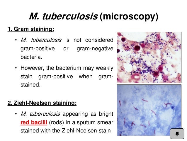
Most of the time, you'll see a mix. Some might appear faintly purple, some might be a bit reddish, and a good chunk of them might just look… confused. It’s like they’re half in, half out of the Gram-positive club. This inconsistency is the first clue that something else is going on here.
The Secret Ingredient: Mycolic Acids
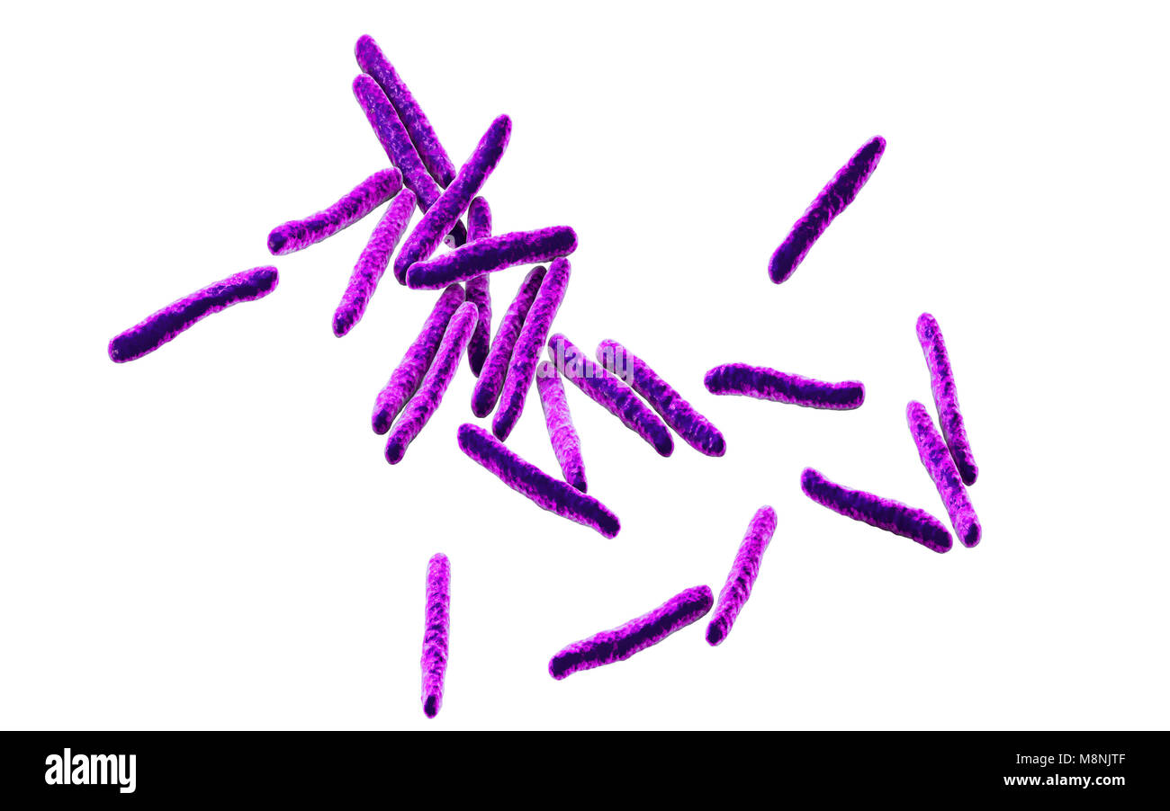
So, what’s their deal? Why are they so indecisive in their Gram staining? The answer, my curious readers, lies in their unique cell wall composition. *Mycobacterium tuberculosis has a significant amount of mycolic acids in its cell wall. And these aren't just any fatty acids; they are waxy, long-chain fatty acids. Think of it as a built-in raincoat, but for bacteria. A really, really effective raincoat.
These mycolic acids form a waxy, hydrophobic layer outside the peptidoglycan layer. This waxy coating is a crucial component of their survival strategy. It makes them resistant to many things that would kill other bacteria, including the harsh conditions within our lungs and, importantly, some of the chemicals used in standard staining procedures.
Because of this waxy layer, the crystal violet stain doesn't penetrate as easily or get as firmly trapped by the peptidoglycan as it does in typical Gram-positive bacteria. And when you introduce the decolorizing alcohol, it can partially strip the stain away. This is why you often see a mix of colors, or very faint staining. It’s not a clear-cut Gram-positive reaction.
The Acid-Fast Stain: The Real Star of the Show
Because the Gram stain isn't the most reliable for identifying Mycobacterium tuberculosis, microbiologists developed a specialized stain: the acid-fast stain. And this, my friends, is where these waxy rebels truly show their colors. The most common type is the Ziehl-Neelsen stain, and it's a bit more… vigorous.
Here's how it generally works: You apply the primary stain (carbolfuchsin) and then, this is the key part, you heat the slide. This heat, combined with the carbolfuchsin, forces the stain to penetrate the waxy mycolic acid layer. Once it's in, it's in. No amount of decolorizing agent (acid-alcohol, in this case, hence "acid-fast") can remove it.
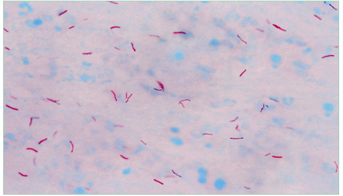
So, what do you see? If the bacteria are acid-fast (and Mycobacterium tuberculosis absolutely is), they will retain the bright red carbolfuchsin stain. Everything else, the background and any non-acid-fast bacteria, will be decolorized and then stained blue by a counterstain (methylene blue). So, the Mycobacterium tuberculosis will stand out as brilliant red rods against a blue background. It’s a much clearer, more definitive result.
Why Does This Matter So Much?
You might be thinking, "Okay, so they're a bit tricky to stain. So what?" Well, in the world of diagnosing and treating infectious diseases, being able to accurately identify the culprit is absolutely paramount. For TB, a timely and accurate diagnosis can literally be the difference between life and death.

The acid-fast stain is often one of the first tests performed when TB is suspected. It provides a quick visual confirmation that Mycobacterium species are present. This allows doctors to start appropriate treatment promptly, even while waiting for more definitive culture results. Imagine if we relied solely on the Gram stain for TB diagnosis; it would be a much less effective and more frustrating process, leading to delays in care.
It's a prime example of how understanding the unique biology of a pathogen – in this case, the waxy cell wall of Mycobacterium – directly impacts our ability to combat it. It's not just about pretty colors under a microscope; it's about saving lives.
The Ironic Twist: Gram-Positive in Spirit, Not in Deed
There’s a certain irony, don’t you think? Mycobacterium tuberculosis has the cellular machinery that would classify it as Gram-positive, yet it consistently behaves like… well, like itself. It’s like having all the ingredients for a chocolate cake but deciding to make a savory pie instead. The basic components are there, but the overall outcome is different.
This is why it’s often described as "gram-variable" or, more accurately, that it stains poorly by the Gram method. It doesn't fit neatly into the binary classification. It forces us to look deeper, to understand the 'why' behind the 'what'. It’s a reminder that nature, and especially the world of microbes, is full of exceptions and complexities.
It makes you wonder, if we just stuck to the textbook definition without any further investigation, how many cases might have been missed or misdiagnosed? This is the beauty and the challenge of scientific inquiry – always questioning, always refining, always seeking a more complete picture.
Beyond the Stain: A World of Resistance
The mycolic acid layer isn't just a nuisance for staining procedures. It’s a fortress. This waxy coating contributes significantly to the bacterium's resistance to:
- Antibiotics: Many drugs have trouble penetrating this barrier, making TB notoriously difficult to treat. Treatment regimens are long and complex, often involving multiple drugs, precisely because of this resistance.
- Drying out: The waxy layer helps the bacteria survive in dry environments, which is crucial for transmission through airborne droplets.
- Host immune responses: It provides protection against the host's immune cells, allowing the bacteria to persist within the body.
So, when we talk about the Gram reaction of Mycobacterium tuberculosis, we're not just talking about a staining artifact. We're talking about a fundamental characteristic that underpins its pathogenicity and its remarkable ability to evade eradication.
In Conclusion: Appreciating the Outliers
So, the next time you hear about a Gram stain, remember that it's not always a black and white (or rather, purple and pink) situation. And especially remember Mycobacterium tuberculosis, the bacterium that makes its own rules and forces us to adapt. It’s a testament to the fact that understanding the nuances, the outliers, the rebels, is often where the most important scientific discoveries are made.
It’s a bit like that one friend who always approaches problems from a completely different angle. Annoying at times, perhaps, but often the one who comes up with the truly innovative solution. Mycobacterium tuberculosis is that friend to the microbiology world. It challenges our assumptions and pushes us to develop better tools and deeper understanding.
And while my initial encounter with it under the microscope might have felt like a battle of wills, it ultimately led to a greater appreciation for the incredible complexity and resilience of life, even at its smallest scale. So, cheers to the waxy, rebellious, and ultimately fascinating Mycobacterium tuberculosis! It might not be a classic Gram-positive, but it’s certainly one to remember.