Normal Chest X Ray Vs Tuberculosis
.png)
Ever found yourself staring at a medical show, or maybe just curious about what’s really going on inside us? Well, buckle up, because we’re about to dive into something that sounds a bit serious but can actually be pretty fascinating: the humble chest X-ray and its connection to a historical, and still very relevant, disease called Tuberculosis. Think of it as a secret detective mission for your lungs!
Why is this Worth Talking About?
It might seem a little strange to call a medical topic "fun," but understanding how we can peek inside our bodies to check for problems is genuinely amazing. A chest X-ray is like a snapshot, a quick look that doctors use to see if everything is ticking along as it should be in your chest. And when we talk about Tuberculosis (often shortened to TB), we're talking about a disease that has shaped human history and is still a significant health concern worldwide. Knowing what a healthy chest looks like on an X-ray, and what signs might point to TB, empowers us with knowledge. It’s like having a little superpower of understanding when it comes to our health!
The Grand Reveal: What Exactly IS a Chest X-ray?
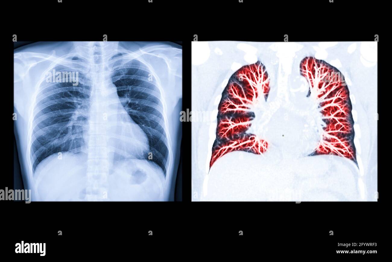
Imagine this: you stand in front of a special machine, maybe wear a lead apron for a little bit of a futuristic vibe, and then – click! – an image of your lungs, heart, and bones appears on a screen or a piece of film. That’s basically a chest X-ray. It uses a tiny amount of radiation, much less than you might think, to create this image. The reason it's so useful is that different parts of your body absorb this radiation differently. Dense structures like bones show up as bright white, while softer tissues like your lungs appear darker. This contrast is what allows doctors to spot abnormalities.
The primary purpose of a chest X-ray is incredibly broad. Doctors use it to:
- Diagnose conditions: Like pneumonia (an infection), fluid in the lungs, or even certain types of cancer.
- Monitor existing conditions: To see how well treatment is working or if a chronic lung disease is progressing.
- Check for injuries: After an accident, to make sure there are no broken ribs or other chest trauma.
- Screen for diseases: Especially in people who might be at higher risk.
The benefits are huge. It’s quick, relatively inexpensive, and doesn’t involve any needles or uncomfortable procedures. It’s a cornerstone of diagnostic medicine, providing a vital first look into the chest cavity.

Enter the Villain (of sorts): Tuberculosis (TB)
Now, let’s talk about Tuberculosis. This is a disease caused by a type of bacteria called Mycobacterium tuberculosis. While it most commonly affects the lungs, TB can also attack other parts of the body, like the kidneys, spine, and brain. For a long time, TB was a dreaded illness, known as "consumption," and it sadly claimed many lives.
Thanks to modern medicine, including powerful antibiotics, TB is treatable and often curable. However, it’s not a thing of the past. It remains a major global health problem, especially in certain regions of the world. This is where our X-ray detective comes in!
The X-ray Detects TB: What to Look For
When a doctor suspects TB, a chest X-ray is often one of the first diagnostic tools. A normal chest X-ray will show clear lungs with no significant shadows or unusual patterns. The airways will be well-defined, and the heart and other structures will appear within their expected size and shape. Think of it as a pristine landscape.

However, in someone with TB, the X-ray might show tell-tale signs. These can include:
White spots or patches: These are often areas where the bacteria have caused inflammation and the body’s immune system is fighting back. They might look like hazy clouds or small cotton balls scattered in the lung tissue.
Cavities: In more advanced cases, the infection can cause the lung tissue to break down, forming empty spaces called cavities. These can appear as dark, hole-like areas on the X-ray.
Hilar lymph node enlargement: The lymph nodes in the center of the chest might appear larger than normal, indicating an immune response.
It's important to remember that these signs are not exclusive to TB. Other conditions, like pneumonia or fungal infections, can sometimes present with similar findings. This is why a radiologist (a doctor who specializes in interpreting medical images) and the patient's overall symptoms are crucial for a proper diagnosis. The X-ray is a piece of the puzzle, not the whole story!
Bringing It All Together
So, while we're not exactly discussing a blockbuster movie plot, the journey from a plain chest X-ray to potentially identifying a disease like TB is a testament to scientific progress and our ability to understand and combat health challenges. A normal chest X-ray is a reassuring sight for doctors and patients alike, showing healthy lungs at work. When abnormalities are present, especially those suggestive of TB, the X-ray serves as a vital clue, guiding further investigation and leading to the necessary treatment to help people recover. It’s a fantastic example of how technology helps us keep our bodies in good working order!